DeLLphi-304
For2L SCLC after platinum, with or without baseline brain metastases
TL;DRPost hoc CNS analysis: median CNS PFS NE vs 7.2mo, HR 0.54 (0.39-0.75) favoring 2L tarlatamab in SCLC.
For an RT reader the actionable number is the brain-met subset: CNS PFS 6.5 vs 4.2mo, HR 0.40 (0.24, 0.66) by mRANO-BM BICR, with CNS CR 14.9% vs 5.4%. But >70% had prior CNS-directed treatment and no RT exposure or intracranial-failure pattern is reported, so this informs surveillance interval rather than deferring SRS.
In relapsed SCLC with treated, asymptomatic brain metastases entering 2L, this supports tarlatamab as systemic therapy with meaningful intracranial activity; it does not address untreated or symptomatic CNS disease, where local therapy remains the studied path.
The brain-met subset read by mRANO-BM BICR gives CNS PFS 6.5 vs 4.2mo, HR 0.40 (0.24, 0.66), which is the number to hold against a systemic-only strategy. But >70% had prior CNS-directed therapy and neither RT exposure nor in-field versus out-of-field failure is reported, so it moves the surveillance interval, not the decision to offer SRS.
Intracranial activity is now a differentiator for tarlatamab in 2L SCLC rather than an unknown: CNS CR 14.9% vs 5.4% and CNS disease control 77.6% vs 71.4%, with median duration of CNS disease control 8.2 vs 5.2mo. That supports keeping a pt with treated, stable brain metastases on the bispecific rather than defaulting to chemotherapy for CNS coverage.

| Arm | n | Median CNS PFS (95% CI) | HR (95% CI) |
|---|---|---|---|
| Tarlatamab | 67 | 6.5 (4.3, 13.7) | 0.40 (0.24, 0.66) |
| Chemotherapy | 56 | 4.2 (2.9, 5.5) | n/a |
+2 more figures

| Arm | n | Median CNS PFS (95% CI) | HR (95% CI) |
|---|---|---|---|
| Tarlatamab | 254 | NE (13.7, NE) | 0.54 (0.39, 0.75) |
| Chemotherapy | 255 | 7.2 (5.6, NE) | n/a |
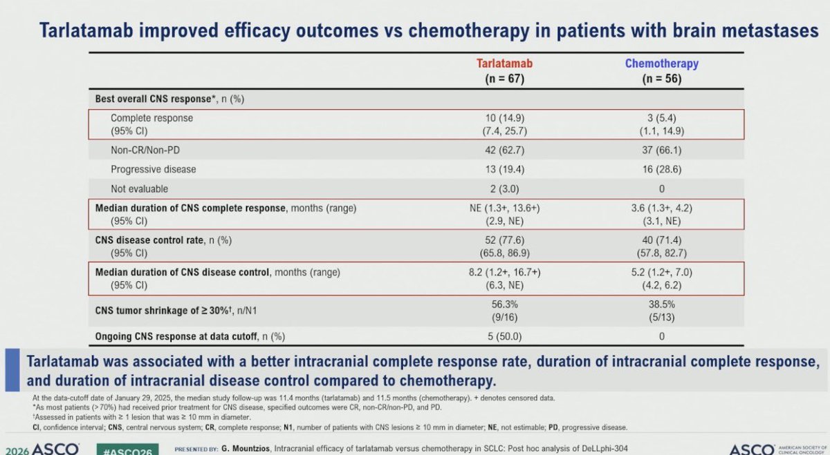
| CNS outcome | Tarlatamab (n=67) | Chemotherapy (n=56) |
|---|---|---|
| Complete response, n (%) | 10 (14.9) | 3 (5.4) |
| Non-CR/non-PD, n (%) | 42 (62.7) | 37 (66.1) |
| Progressive disease, n (%) | 13 (19.4) | 16 (28.6) |
| CNS disease control rate, n (%) | 52 (77.6) | 40 (71.4) |
| Median duration of CNS disease control, mo | 8.2 (1.2+, 16.7+) | 5.2 (1.2+, 7.0) |
10 details 1 trial watching
Post hoc intracranial analysis of the randomised DeLLphi-304 trial of second-line tarlatamab versus chemotherapy in SCLC. Two assessment frameworks: investigator RECIST in the ITT population and mRANO-BM by BICR in pts with baseline brain metastases. Data cutoff January 29, 2025; median follow-up 11.4 mo (tarlatamab) and 11.5 mo (chemotherapy).
ITT n=254 tarlatamab versus n=255 chemotherapy. The brain-metastasis cohort was n=67 versus n=56, defined as ≥1 brain metastasis at baseline per mRANO-BM by BICR plus ≥1 post-baseline scan. More than 70% had already received CNS-directed treatment, which is why the response categories were CR, non-CR/non-PD and PD rather than a conventional ORR.
This analysis reports time to CNS progression or death (CNS PFS) in the ITT population and in the brain-metastasis subset, plus best overall CNS response, CNS disease control rate, duration of CNS complete response and duration of CNS disease control. None of these was the trial's registered primary endpoint.
Both CNS PFS comparisons favor tarlatamab, and the effect is larger in the brain-metastasis subset (HR 0.40) than in the ITT population (HR 0.54). Intracranial complete response was 14.9% versus 5.4%, and CNS tumor shrinkage ≥30% occurred in 9/16 versus 5/13 pts with measurable lesions ≥10mm.
Brain metastases develop in the majority of SCLC pts, and second-line systemic options have historically been judged on extracranial disease with CNS control assumed to be the province of SRS, WBRT or prophylactic cranial irradiation. A bispecific T-cell engager showing an intracranial complete response rate of 14.9% is the notable part, because the working assumption for large-molecule agents has been limited intracranial penetration.
The ITT median CNS PFS was not estimable at 11.4 months of follow-up, so the ITT curve rests on the early portion of the data and the point estimate can still move. The two populations were also read with different tools and different models (stratified Cox for ITT, unstratified for the subset), so the ITT and subset HRs are not strictly comparable to each other.
The result establishes that intracranial disease does not progress faster under tarlatamab than under chemotherapy, and probably progresses more slowly. What it does not establish is whether that activity is sufficient to defer local therapy in a patient who would otherwise be referred for SRS, since the source reports no radiotherapy exposure data and no in-field versus out-of-field failure pattern.
Post hoc intracranial analysis of a randomised trial; CNS endpoints were not the registered primary, and the brain-met subset HR comes from an unstratified model.
- Does intracranial activity permit deferral of SRS in untreated brain mets
- Activity in CNS-treatment-naive or symptomatic brain metastases n=35 · primary completion 2029-02 · phase 2 tarlatamab, active asymptomatic brain mets
- Durability of CNS control beyond 12 months follow-up
📚 Sources · 🐦 1 tweet
Dr. @g_mountzios #ASCO26 presents CNS outcomes with 2L tarlatamab in DeLLphi-304. Improved time to CNS progression overall (HR 0.54). In pts with brain nets, tarlatamab vs chemo CNS CR rate 15% vs 5% with DCR 78% vs 71% and time to CBS progression 6.5m vs 4.2m, HR 0.40 pic.twitter.com/5i8jL1zlKW
— Stephen V Liu, MD (@StephenVLiu) June 1, 2026
The longer read
The reason this post hoc analysis is worth a radiation oncologist's attention is not the direction of the result, which is unsurprising for an agent that outperformed chemotherapy on the trial's main endpoints, but the intracranial complete response rate. A CNS CR of 14.9% versus 5.4% for a bispecific T-cell engager cuts against the default assumption that a large molecule contributes little inside the CNS, and if it holds it changes how the modality is positioned relative to local therapy rather than merely adding another active systemic option.
The magnitude has to be read with the population in mind. More than 70% of the brain-metastasis cohort had already received CNS-directed treatment, and that fact is doing a lot of work in both directions. It is why the analysis could not report a conventional intracranial ORR and had to fall back on CR, non-CR/non-PD and PD categories, and it means the intracranial disease being followed is largely previously irradiated disease, where the observed benefit could plausibly reflect delayed regrowth of treated lesions, control of new lesions, or both. Those are clinically different things and this readout cannot separate them. A patient with untreated brain metastases is not the patient studied here.
On methodology, two features should move confidence in opposite directions. Favorably, the brain-metastasis analysis used mRANO-BM read by blinded independent central review rather than investigator assessment, which is the right instrument for intracranial disease and the harder standard. Unfavorably, this was post hoc, the CNS endpoints were not the registered primary, and the subset hazard ratio came from an unstratified Cox model while the ITT estimate used a stratified one. That the subset effect (HR 0.40) is larger than the ITT effect (HR 0.54) is the kind of pattern that can be real, since the ITT population dilutes the signal with pts who have no CNS disease to protect, but it is also the pattern that subgroup analyses produce by chance. The confidence interval, 0.24 to 0.66, is wide.
Follow-up is the other constraint. At a median of 11.4 and 11.5 months the ITT median CNS PFS was not estimable, with a lower bound of 13.7 months, so the ITT comparison is anchored on the early part of the curve and the eventual point estimate is unsettled. Duration data carry the same caveat: median duration of CNS disease control was 8.2 months versus 5.2 months, both with censored observations at the upper end.
What this does not settle is the decision most likely to arise in clinic. Nothing here reports radiotherapy exposure during the trial, whether pts received SRS on study, or how intracranial failures distributed between previously treated and new sites. Without that, the analysis supports treating tarlatamab as a systemic agent with genuine intracranial activity, and supports a surveillance imaging strategy that does not assume rapid CNS escape, but it does not support deferring local therapy in a patient who would otherwise be referred for it. The trial that would settle that question has not been run.