onc brain

About · curated by Nick Boehling, MD · @nb2276

2026-06-01 ASCO Annual Meeting 2026

digest generated 2026-08-11

DeLLphi-304 post hoc: 2L tarlatamab CNS PFS NE vs 7.2mo (HR 0.54), 6.5 vs 4.2mo in brain-met subset (HR 0.40).
Thoracic carried the day on a CNS question radiation oncologists own: tarlatamab delayed intracranial progression in 2L SCLC, but with no RT exposure data the SRS/WBRT deferral question stays open. Lower GI adds a ctDNA surveillance caution, 41% sensitivity for local regrowth is too low to relax nonoperative imaging.

GI Lower

Watch-and-wait surveillance after chemoRT: ctDNA misses most local regrowths, so endoscopy and MRI keep carrying the salvage window.

Caveats dominate

ctDNA and Local Regrowth/Distant Mets in Nonoperative Rectal Cancer

ForMSS stage I-III rectal ca, cCR/nCR after NAT, on nonoperative management

TL;DRctDNA sensitivity for local regrowth only 41% (12/29), specificity 94%; distant mets sensitivity 74%, specificity 97%.

Why it mattersRadiation oncology

The number that gates watch-and-wait practice is 41% sensitivity for local regrowth (12/29 samples): a negative ctDNA cannot license lengthening MRI or endoscopy intervals in an organ-preservation protocol. Specificity 94% means a positive result is worth acting on, and ctDNA+ at regrowth tracked ypT3-4 in 6/8 vs 3/14 (p=0.01).

Monday clinic

In a stage I-III MSS rectal pt in watch-and-wait after TNT or CRT, a positive ctDNA supports intensified assessment for regrowth or distant disease; a negative result does not support relaxing endoscopic or MRI surveillance intervals.

The longer read
ctDNA and Local Regrowth/Distant Mets in Nonoperative Rectal Cancer
OutcomeSensitivitySpecificityAccuracy
For local regrowth12 / 29 (41%)480 / 509 (94%)492 / 538 (91%)
For distant metastasis31 / 42 (74%)611 / 627 (97%)642 / 669 (96%)
11 details

Single-institution cohort from MD Anderson's INTERCEPT program, 2020-2024, N=110, with serial tumor-informed ctDNA during nonoperative management. Median follow-up 25 months (IQR 18-37).

Microsatellite stable stage I-III rectal adenocarcinoma achieving cCR or near-cCR after neoadjuvant therapy and managed nonoperatively. Median age 56; baseline cT3 in 72 and cT4 in 16; 69 received TNT, 41 CRT or chemotherapy alone.

Local regrowth and distant metastasis by longitudinal ctDNA status, by first post-NAT ctDNA (within 180 days), and per-sample accuracy for an event within ±90 days of each draw. Salvage-surgery pathology by ctDNA status at regrowth.

Twenty-three pts (21%) had local regrowth and 12 (11%) distant metastasis. Ever-positive ctDNA separated both curves (log rank p=0.0002 for regrowth, p<.0001 for metastasis). The per-sample table carries the operating characteristics.

MSS stage I-III rectal cancer in cCR/nCR on nonoperative management with tumor-informed ctDNA surveillance
Does not represent MSI-high disease, pts taken to upfront TME, or ctDNA assays other than the tumor-informed exome platform used here.

Per-sample analysis pools 669 draws from 110 pts without accounting for repeated measures, so the confidence around 41% is softer than the denominator suggests. The first-post-NAT comparison rests on n=12 evaluable pts as reported in the source. No comparison against MRI or endoscopy, the tests ctDNA would have to beat.

The asymmetry between local (41%) and distant (74%) sensitivity is the informative result: intraluminal regrowth from a small residual burden sheds too little DNA to be caught reliably, while metastatic disease does. That biology argues for ctDNA as a distant-recurrence tool layered onto, not substituted for, luminal surveillance.

Retrospective single-center cohort; per-sample analysis treats 669 draws from 110 pts as independent. No head-to-head against MRI/endoscopy surveillance.

  • Does ctDNA add anything over MRI plus endoscopy in NOM surveillance?
  • Can draw timing or a lower assay threshold raise local regrowth sensitivity?
  • Does ctDNA-triggered restaging improve salvage outcomes?

Sourced from @NiuSanford

📚 Sources · 🐦 1 tweet

Thoracic / Lung

Post hoc intracranial read on a systemic agent, framed against the SRS/WBRT timing decision it does not yet resolve.

Caveats dominate

DeLLphi-304

For2L SCLC after platinum, with or without baseline brain metastases

TL;DRPost hoc CNS analysis: median CNS PFS NE vs 7.2mo, HR 0.54 (0.39-0.75) favoring 2L tarlatamab in SCLC.

Why it mattersRadiation oncology

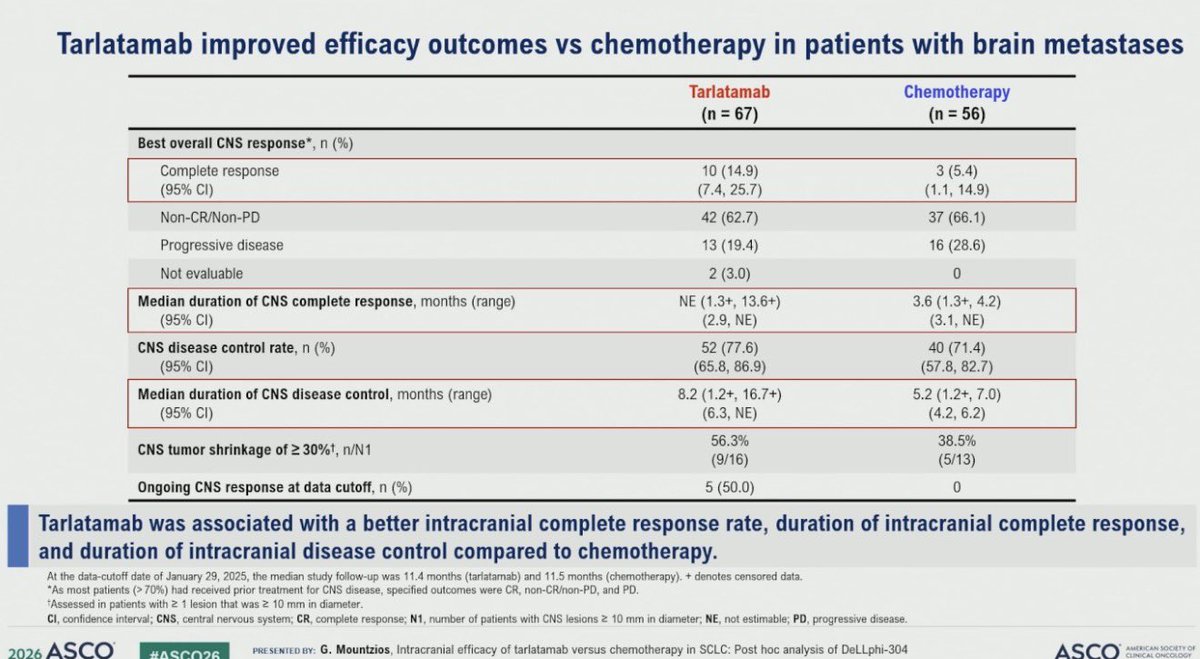
For an RT reader the actionable number is the brain-met subset: CNS PFS 6.5 vs 4.2mo, HR 0.40 (0.24, 0.66) by mRANO-BM BICR, with CNS CR 14.9% vs 5.4%. But >70% had prior CNS-directed treatment and no RT exposure or intracranial-failure pattern is reported, so this informs surveillance interval rather than deferring SRS.

Monday clinic

In relapsed SCLC with treated, asymptomatic brain metastases entering 2L, this supports tarlatamab as systemic therapy with meaningful intracranial activity; it does not address untreated or symptomatic CNS disease, where local therapy remains the studied path.

The longer read
DeLLphi-304
ArmnMedian CNS PFS (95% CI)HR (95% CI)
Tarlatamab676.5 (4.3, 13.7)0.40 (0.24, 0.66)
Chemotherapy564.2 (2.9, 5.5)n/a
+2 more figures
DeLLphi-304
ArmnMedian CNS PFS (95% CI)HR (95% CI)
Tarlatamab254NE (13.7, NE)0.54 (0.39, 0.75)
Chemotherapy2557.2 (5.6, NE)n/a
DeLLphi-304
CNS outcomeTarlatamab (n=67)Chemotherapy (n=56)
Complete response, n (%)10 (14.9)3 (5.4)
Non-CR/non-PD, n (%)42 (62.7)37 (66.1)
Progressive disease, n (%)13 (19.4)16 (28.6)
CNS disease control rate, n (%)52 (77.6)40 (71.4)
Median duration of CNS disease control, mo8.2 (1.2+, 16.7+)5.2 (1.2+, 7.0)
10 details 1 trial watching

Post hoc intracranial analysis of the randomised DeLLphi-304 trial of second-line tarlatamab versus chemotherapy in SCLC. Two assessment frameworks: investigator RECIST in the ITT population and mRANO-BM by BICR in pts with baseline brain metastases. Data cutoff January 29, 2025; median follow-up 11.4 mo (tarlatamab) and 11.5 mo (chemotherapy).

ITT n=254 tarlatamab versus n=255 chemotherapy. The brain-metastasis cohort was n=67 versus n=56, defined as ≥1 brain metastasis at baseline per mRANO-BM by BICR plus ≥1 post-baseline scan. More than 70% had already received CNS-directed treatment, which is why the response categories were CR, non-CR/non-PD and PD rather than a conventional ORR.

This analysis reports time to CNS progression or death (CNS PFS) in the ITT population and in the brain-metastasis subset, plus best overall CNS response, CNS disease control rate, duration of CNS complete response and duration of CNS disease control. None of these was the trial's registered primary endpoint.

Both CNS PFS comparisons favor tarlatamab, and the effect is larger in the brain-metastasis subset (HR 0.40) than in the ITT population (HR 0.54). Intracranial complete response was 14.9% versus 5.4%, and CNS tumor shrinkage ≥30% occurred in 9/16 versus 5/13 pts with measurable lesions ≥10mm.

Brain metastases develop in the majority of SCLC pts, and second-line systemic options have historically been judged on extracranial disease with CNS control assumed to be the province of SRS, WBRT or prophylactic cranial irradiation. A bispecific T-cell engager showing an intracranial complete response rate of 14.9% is the notable part, because the working assumption for large-molecule agents has been limited intracranial penetration.

relapsed SCLC entering second line, including pts with baseline brain metastases the majority of whom had prior CNS-directed therapy
Does not represent untreated, symptomatic or leptomeningeal CNS disease, which this analysis does not report on.

The ITT median CNS PFS was not estimable at 11.4 months of follow-up, so the ITT curve rests on the early portion of the data and the point estimate can still move. The two populations were also read with different tools and different models (stratified Cox for ITT, unstratified for the subset), so the ITT and subset HRs are not strictly comparable to each other.

The result establishes that intracranial disease does not progress faster under tarlatamab than under chemotherapy, and probably progresses more slowly. What it does not establish is whether that activity is sufficient to defer local therapy in a patient who would otherwise be referred for SRS, since the source reports no radiotherapy exposure data and no in-field versus out-of-field failure pattern.

Post hoc intracranial analysis of a randomised trial; CNS endpoints were not the registered primary, and the brain-met subset HR comes from an unstratified model.

  • Does intracranial activity permit deferral of SRS in untreated brain mets
  • Activity in CNS-treatment-naive or symptomatic brain metastases
    n=35 · primary completion 2029-02 · phase 2 tarlatamab, active asymptomatic brain mets
  • Durability of CNS control beyond 12 months follow-up

Sourced from @StephenVLiu

📚 Sources · 🐦 1 tweet