CNS
2026-07-13
EANO Consensus Statement on Radiation Necrosis
ForPost-cranial-RT pts with a new enhancing lesion, glioma or brain metastases
TL;DRDelphi consensus: 53/57 statements reached ≥80% agreement across 20 experts; perfusion MRI + amino acid PET preferred, bevacizumab for steroid-refractory RN.
The dose-volume numbers are the transferable part: V12Gy ~10 cm³ carrying 5-10% symptomatic necrosis in single-fraction SRS, and hypofractionated brain+target V20Gy (3fx) or V24Gy (5fx) <20 cm³ holding RN needing resection under 4%. That is a planning constraint, and it gates the single-session vs hypofractionated choice for larger or eloquent-region metastases.
For a patient 6-24 months out from SRS or cranial RT with a new enhancing lesion, this supports going to perfusion MRI plus amino acid PET before calling progression; it does not settle mixed lesions, where the panel still points to tissue.
The planning-relevant numbers: V12Gy ~10 cm³ carries 5-10% symptomatic necrosis after single-fraction SRS, while hypofractionated brain+target V20Gy (3fx) or V24Gy (5fx) <20 cm³ keeps RN requiring resection under 4%. Re-irradiation risk climbs to 25% above cumulative EQD2 130 Gy, which constrains repeat SRS for lesions over 2.0-3.0 cm.
Bevacizumab moves from rescue to a triggered step at steroid dependence >4 weeks or dexamethasone 8 mg/day, with four accepted dosing schemes and no evidence favouring one. The panel also flags added RN risk when SRS is combined with dual checkpoint blockade or antibody-drug conjugates, which bears on sequencing brain RT against systemic therapy.
Resection carries the lowest accepted agreement in the management table (80.0%) yet is positioned as giving diagnosis and definitive treatment in one step for accessible lesions. LITT reached 100% agreement as the option for treatment-resistant RN unsuitable for resection, which gives a documented pathway for the steroid-dependent or bevacizumab-cycling patient.
11 details 5 trials watching
Three-round Delphi. 57 statements drafted after literature review across epidemiology-pathophysiology (n=8), causes (n=10), imaging (n=10), histopathology (n=9), management (n=20), plus an exploratory research set of 8. Consensus prespecified at ≥80% agreement on a 5-point Likert scale.
The voting panel, not patients: 15 volunteers (2 radiation/clinical oncologists, 4 neuro-oncologists, 4 medical oncologists, 2 neurosurgeons, 1 neuropathologist, 1 neuroradiologist, 1 nuclear medicine physician), expanded by 5 more in round 1 (4 neuro-oncologists, 1 radiation/clinical oncologist) to 20 total. Clinically the scope is RN after RT for primary glial or metastatic brain tumors.
RN is called uncommon below EQD2 ~40 Gy (α/β 2 Gy), rising with dose per fraction, total dose and irradiated volume. V12Gy ~10 cm³ carries a 5-10% symptomatic necrosis risk after single-session SRS; for hypofractionated SRT, brain-plus-target V20Gy (3fx) or V24Gy (5fx) <20 cm³ associates with <10% any necrosis and <4% RN requiring resection. Re-irradiation risk stays under 10% at cumulative EQD2 100-110 Gy and reaches up to 25% above 130 Gy.
Consensus reached on 53 of 57 statements after three rounds. Diagnostic and management statements are tabulated separately in the details; the four that failed to reach 80% are not identified in the available text.
| Modality | Setting | Sensitivity | Specificity |
|---|---|---|---|
| T1 contrast-enhanced MRI alone | WHO grade 3-4 glioma | 68% | 77% |
| T1 contrast-enhanced MRI alone | Brain metastases | 79% | 76% |
| DSC rCBV | Grade 3-4 glioma | 87-90% | 86-88% |
| DSC rCBV | Brain metastases (418 lesions) | 83% | 78% |
| MR spectroscopy Cho/NAA | Glioma (455 pts) | 88% | 86% |
| Amino acid PET | Brain metastases (~400 pts) | 82% | 84% |
The document's practical contribution is a decision sequence rather than a new treatment: advanced MRI first, amino acid PET to add confidence, tissue when doubt persists, and treatment triggered only by symptoms of >7 days with increasing severity. Its most consequential position is moving bevacizumab from rescue to an expected next step at a defined steroid threshold (dependence >4 weeks or dexamethasone 8 mg/day), which is a lower bar than many centres apply.
Agreement percentages measure how a self-selected 20-person European panel voted, and the panel skews neuro-oncology (8 of 20) over radiation oncology (3 of 20) for a complication defined by dose and volume. FET and F-DOPA availability is largely European, so the imaging algorithm's top tier is not reproducible in most centres worldwide. The 4 non-consensus statements are not enumerated in the accessible text, so the reader cannot see where the panel actually split.
Delphi expert-opinion document, no efficacy endpoint. Agreement measured against opinion, not outcomes; authors state absence of Level 1 evidence and call for randomised trials.
- Optimal bevacizumab dose, interval and duration for symptomatic RN n=408 · primary completion 2028-07 · phase 3 bevacizumab vs dexamethasone, 1L sCRNrecruiting Corticodependent or Corticoresistant Brain Radionecrosis After Radiotherapy for Brain Metastases Phase 3n=84 · primary completion 2028-08 · randomised bev vs placebo in steroid-refractory RN
- Single-session SRS vs hypofractionated SRT for RN risk active Comparing Single vs Multiple Dose Radiation for Cancer Patients With Brain Metastasis and Receiving Immunotherapy Phase NAn=58 · primary completion 2028-03 · single vs fractionated SRS toxicity on ICIn=54 · primary completion 2029-02 · staged SRS vs FSRT, 2-5 cm mets, safety endpointn=504 · primary completion 2030-01 · phase 3 3x10 Gy SRT vs 1x20-25 Gy SRS
- Predictors of bevacizumab response and recurrence rate after treatment
📚 Sources · 📄 1 paper
Abstract
2026-07-07
EORTC 22033-26033/NCIC-CTG/TROG/MRC-CTU
ForClinical high-risk WHO grade 2 glioma, first-line, molecularly classified
No significant difference
PFS and OS both ns between RT and TMZ arms; effect size not reported in source
TL;DRMature phase III: RT (50.4Gy/28fx) vs dose-dense TMZ in high-risk WHO grade 2 LGG, no PFS or OS difference in any molecular subtype.
The RT-relevant read is what this does NOT license: single-modality TMZ never beat RT, so deferring 50.4Gy/28fx to spare late toxicity buys nothing on PFS or OS. IDH wild-type was the one split (OS 2.5 vs 4.7 yrs, HR 0.47 [0.27-0.82], P=.0068), post hoc and n=64, and those tumors now grade as GBM.
In molecularly classified high-risk WHO grade 2 glioma being planned for first-line therapy, this supports neither leading modality over the other and does not address the combined-modality RT plus alkylator approach that is standard for IDH-mutant astrocytoma.
50.4Gy in 28fx as sole therapy was not beaten by TMZ, so deferring RT to postpone late neurocognitive cost does not buy an outcome advantage. The trial says nothing about RT dose, target volume or the combined-modality regimen a high-risk grade 2 astrocytoma now receives, so it does not move the modern RT decision, only closes the omission argument.
Dose-dense TMZ 75 mg/m² 21/28d for up to 12 cycles as sole first-line therapy is equivalent, not superior, to RT, and the schedule was never tested against the combined-modality standard. The IDH wild-type OS advantage (2.5 vs 4.7 yrs, HR 0.47 [0.27-0.82]) is post hoc in n=64 tumors that now classify as GBM.
8 details 2 trials watching
Randomised phase III, four-group intergroup trial (EORTC/NCIC-CTG/TROG/MRC-CTU), N=478, comparing two single-modality first-line strategies. This is the mature analysis; a post hoc reclassification to 2021 WHO criteria was possible in 73% (351/478) with analyzable tissue.
Clinical high-risk WHO grade 2 low-grade glioma, treatment-naive. Post hoc molecular strata: IDHmt/1p19q non-codeleted astrocytoma n=178, IDHmt/1p19q codeleted oligodendroglioma n=109, IDH wild-type n=64.
Standard fractionation, 28 × 1.8 Gy (50.4 Gy), delivered as the entire assigned treatment. No concurrent or adjuvant systemic therapy in the RT arm, which is the gap between this trial and how grade 2 glioma is treated now.
Dose-dense temozolomide 75 mg/m² once daily, 21 of 28 days, up to 12 cycles, given as sole first-line therapy with RT withheld.
Primary: progression-free survival. Overall survival and differential response by molecular marker were the mature-analysis questions.
No significant difference in PFS or OS between arms, and the null held across every molecular subtype except IDH wild-type, where OS favored TMZ.
| Subtype (n) | RT | TMZ | HR |
|---|---|---|---|
| IDHmt astrocytoma, non-codel (n=178) | 6.6-6.7 yrs (either arm) | 6.6-6.7 yrs (either arm) | 0.67-1.44, P=.93 |
| IDHmt oligodendroglioma, 1p/19q codel (n=109) | 12.9 yrs (9.4-NR) | 14.9 yrs (10.1-NR) | 0.88 (0.52-1.49), P=.63 |
| IDH wild-type (n=64) | 2.5 yrs (1.8-3.3) | 4.7 yrs (2.2-7.2) | 0.47 (0.27-0.82), P=.0068 |
RTOG 9802 established RT followed by PCV as the high-risk grade 2 benchmark, and combined-modality therapy is now standard for IDH-mutant astrocytoma. EORTC 22033 tested neither arm of that question: it asked which single modality to lead with, and answered that it does not matter.
Molecular strata are post hoc in 73% of the enrolled population, so subtype comparisons carry both selection and multiplicity risk. The IDH wild-type finding rests on n=64, and under 2021 criteria those tumors would not be enrolled as grade 2 at all.
The durable contribution is prognostic rather than therapeutic: median OS of 12.9-14.9 years in codeleted oligodendroglioma against 6.6-6.7 years in IDH-mutant astrocytoma quantifies how far molecular class outweighs modality choice. The finding that pts ≥40 fared better than <40 undercuts the age-40 cutoff that still gates high-risk definitions.
Mature randomised phase III, primary endpoint negative in both arms and across molecular subtypes; the IDHwt survival difference is post hoc, n=64.
- Optimal sequencing of RT and alkylator in IDH-mutant astrocytoma
- Whether IDH inhibitors displace upfront RT in low-risk grade 2 glioma n=90 · primary completion 2027-10 · PROs: vorasidenib vs RT vs surveillance in grade 2 IDH-mutn=150 · primary completion 2031-11 · real-world vorasidenib, grade 2, no immediate RT need
- Late neurocognitive cost of combined-modality vs single-modality
📚 Sources · 📄 1 paper
Abstract
2026-06-09
NRG Oncology RTOG 0539 NCT00895622
ForWHO grade 1-3 meningioma, newly diagnosed or recurrent, any resection extent
TL;DR10-yr PFS 85.2% observed low-risk, 72.2% intermediate-risk at 54 Gy, 42.5% high-risk at 60 Gy.
The transferable RT read is the target: 54 Gy/30 fx for intermediate-risk and 60 Gy/30 fx for high-risk, with grade 3+ RT-attributed toxicity 9.6% and 15.1%. Recurrent grade 1 salvaged with RT reached only 67.0% 10-yr OS, worse than upfront grade 2 post-GTR at 91.0%, which argues against deferring RT in a grade 1 you expect to recur.
In newly diagnosed WHO grade 2 meningioma after GTR, this supports upfront 54 Gy while NRG BN003 and ROAM read out; it does not speak to observation in that group, since no untreated grade 2 arm was enrolled.
The transferable parameters are 54 Gy/30 fx intermediate-risk and 60 Gy/30 fx high-risk, with grade 3+ RT-attributed toxicity of 9.6% and 15.1%. Recurrent grade 1 salvaged with RT reached only 67.0% 10-yr OS vs 91.0% for upfront grade 2 post-GTR, which argues against deferring RT in a grade 1 likely to recur.
Extent of resection carried more weight than histology: STR vs GTR gave PFS HR 2.58 (1.09-6.11) and OS HR 3.38 (1.28-8.91) on multivariable analysis. Even in observed low-risk grade 1 disease, 10-yr PFS was 88.0% after GTR vs 72.7% after STR, so a Simpson-grade decision at the first operation still shows at 10 years.
10 details 4 trials watching
Multi-arm prospective phase 2 trial (NCT00895622) of risk-adapted management, not randomised: each risk group followed its own assigned strategy. 244 consented, 165 eligible and treated per protocol. Original primary endpoint was 3-yr PFS, previously reported; this is the mature analysis with data cutoff 8/15/2023 and median follow-up 12.1, 12.0 and 11.1 years across the three cohorts.
Adults ≥18 with Zubrod 0-1 and histologically confirmed unifocal WHO grade 1-3 meningioma, newly diagnosed or recurrent, any resection extent, with centrally confirmed pathology. Median age 56, 62 in the high-risk group; 65.5% female overall. Recurrent disease made up 30.8% of the intermediate and 47.2% of the high-risk cohorts.
Group 1 (grade 1 post-GTR/STR) was observed only. Group 2 (recurrent grade 1, or newly diagnosed grade 2 post-GTR) received 54 Gy in 30 fractions. Group 3 (newly diagnosed grade 2 post-STR, newly diagnosed grade 3, or recurrent grade 2/3) received 60 Gy in 30 fractions.
The gradient tracks risk assignment cleanly at 10 years, and the two Cox covariates that survive adjustment are recurrent disease and subtotal resection, both for PFS and OS. See the cohort and covariate tables above.
| Cohort | Management | 10-yr PFS | 10-yr OS | 10-yr cum. incidence progression |
|---|---|---|---|---|
| Low (grp 1, n=60) | Observation | 85.2% (75.7-94.8) | 94.1% (87.6-100) | 8.9% (3.2-18.2) |
| Intermediate (grp 2, n=52) | RT 54 Gy | 72.2% (59.2-85.1) | 84.7% (74.2-95.2) | 21.2% (10.8-33.9) |
| High (grp 3, n=53) | RT 60 Gy | 42.5% (28.7-56.3) | 51.1% (37.0-65.2) | 39.3% (25.8-52.5) |
| Covariate | PFS HR (95% CI), p | OS HR (95% CI), p |
|---|---|---|
| Recurrent vs initial | 2.5 (1.01-6.18), p=0.047 | 2.86 (1.06-7.70), p=0.038 |
| STR vs GTR | 2.58 (1.09-6.11), p=0.031 | 3.38 (1.28-8.91), p=0.014 |
Grade 3+ AEs attributed to radiotherapy occurred in 5 pts (9.6%) of the intermediate-risk and 8 pts (15.1%) of the high-risk cohorts. Newly reported late events in the intermediate group were auditory and neurologic grade 3 plus one grade 4 hemorrhage. Zubrod, MMSE and neurologic function score were stable over time.
This is the mature counterpart to the trial's own 3-yr landmark reports and now sits as the benchmark alongside the ongoing de-escalation questions in NRG BN003 (NCT03180268) and ROAM/EORTC 1308, both of which test whether grade 2 post-GTR needs RT at all. Until those read out, the 10-yr PFS 72.2% and OS 84.7% here are the reference numbers for treating that group.
Pathology was graded under the WHO criteria of the enrolment era, so some group 1 tumors would likely be reclassified today, which is the trial's own proposed explanation for the poor recurrent grade 1 outcomes. Subgroup estimates rest on very small denominators, with intervals such as 15.0% (0-42.0%) for recurrent grade 2 PFS that cannot support a practice decision on their own.
The trial settles the low-risk question (observe after GTR, 10-yr PFS 88.0%) and confirms that high-risk disease is not controlled by 60 Gy, with 10-yr PFS 42.5%. What it cannot settle is whether the intermediate-risk result reflects RT or favorable biology, since no group 2 patient went untreated.
Non-randomised risk-adapted phase 2 with mature 10+ yr follow-up; supports existing consensus (observe post-GTR grade 1, RT otherwise) rather than testing it against a control.
- Does grade 2 meningioma post-GTR need adjuvant RT at all recruiting Observation or Radiation Therapy in Treating Patients With Newly Diagnosed Grade II Meningioma That Has Been Completely Removed by Surgery Phase 3n=163 · primary completion 2027-06 · randomised RT vs observation, GTR grade II
- Whether molecular classification reassigns recurrent grade 1 tumors n=210 · primary completion 2026-03 · methylation + histone PTM signature for recurrence
- How to improve first-line treatment for high-risk meningioma recruiting Vismodegib, FAK Inhibitor GSK2256098, Capivasertib, and Abemaciclib in Treating Patients With Progressive Meningiomas Phase 2n=124 · primary completion 2027-01 · mutation-matched targeted arms, progressive tumorsactive A Trial of Increased Dose Intensity Modulated Proton Therapy (IMPT) for High-Grade Meningiomas Phase NAn=21 · primary completion 2027-08 · dose-escalated IMPT, STR grade II / grade III
📚 Sources · 📄 1 paper
Abstract
FIRESTORM
ForHigh-risk meningioma (WHO gr 2 post-STR, recurrent gr 2, any gr 3), postop RT
HR 0.40
95% CI 0.24-0.69, P = .001 (MVA); IPTW HR 0.45 (0.24-0.83)
TL;DR5-yr PFS 65.8% vs 38.8% with dose-escalated postop RT (BED ≥79.2 Gy) in high-risk meningioma; MVA HR 0.40.
The benefit survives the obvious confounders: excluding the 35 single-fraction SRS cases from SD-RT, DE-RT still gave 5-yr PFS 65.8% vs 41.7% (HR 0.56, 0.36-0.86), and photon-alone escalation matched carbon on PFS (68.3% vs 61.1%) with 0% grade ≥3 radionecrosis. That makes 66-70 Gy conventionally fractionated photons the practical escalation route, not particle referral.
In a subtotally resected WHO grade 2 or any grade 3 meningioma being planned for postoperative RT, this supports considering escalation beyond 60 Gy/30 fx rather than defaulting to it; it does not speak to gross-totally resected grade 1 disease.
Photon-alone escalation to 70 Gy/35 fx or 66 Gy/33 fx matched carbon on 5-yr PFS (68.3% vs 61.1%) with zero grade ≥3 radionecrosis, versus 9.1% in the carbon subgroup. Boost PTV margins were 0.5 to 2 mm off cavity and residual tumor. That makes escalation a standard photon department decision, not a particle referral.
14 details
Individual patient-level meta-analysis pooling 248 patients from 7 international institutions, all retrospective except one prospective trial. Median follow-up 67.1 months (range 2.13-178). PFS was the primary outcome, analyzed by Kaplan-Meier, Cox MVA, and IPTW propensity weighting.
High-risk meningioma per RTOG 0539: newly diagnosed WHO grade 2 after STR/biopsy, any recurrent grade 2, or any grade 3. 188 (75.8%) were grade 2, 103 (41.5%) recurrent, and 182 (75.2%) had Simpson grade 4/STR. Median age 60; 55 (22.2%) had prior RT.
DE-RT was defined by biologically effective dose ≥79.2 Gy (equivalent to 66 Gy in 33 fractions) or receipt of a carbon-ion boost; anything below that threshold counted as SD-RT. Photon DE-RT used a sequential or simultaneous integrated boost with a 0.5 to 2 mm PTV margin, versus 1-2 cm CTV margins in the SD-RT arm. Carbon DE-RT boosted 16 Gy/8 fx after 50 Gy/25 fx photons with a 6 mm CTV margin.
Whole-cohort 3- and 5-yr PFS were 62.8% and 45.0%. DE-RT improved 3-yr (86.4% vs 55.6%) and 5-yr PFS (65.8% vs 38.8%), P = .0022, holding on stratified Cox by grade (HR 0.40, 0.22-0.73) and after IPTW (3-yr 84.7% vs 55.8%). OS was not improved on MVA or IPTW.
| Subgroup | 3-yr PFS DE-RT vs SD-RT | 5-yr PFS DE-RT vs SD-RT | MVA HR (95% CI) | P |
|---|---|---|---|---|
| Simpson 1-3 | 87.5% vs 55.9% | 70.0% vs 40.0% | 0.31 (0.08-1.14) | .08 |
| Simpson 4-5 | 86.3% vs 55.4% | 63.3% vs 38.4% | 0.55 (0.36-0.84) | .006 |
| Cohort | Any-grade RN | Grade ≥3 RN |
|---|---|---|
| DE-RT overall | 20 of 59 (33.9%) | 3 of 59 (5.1%) |
| DE-RT mixed carbon/photon | 15 of 33 (45%) | 3 of 33 (9.1%) |
| DE-RT photon-alone | 5 of 26 (19.2%) | 0% |
| SD-RT | 25 of 189 (13.2%) | 6 of 189 (3.2%) |
Any-grade radionecrosis was 33.9% with DE-RT vs 13.2% with SD-RT (P = .001), but grade ≥3 RN did not differ (5.1% vs 3.2%, P = .47). All 3 grade ≥3 events in the DE-RT arm occurred in the mixed carbon-photon cohort; the photon-alone DE-RT cohort had none, and its any-grade RN rate was not significantly higher than SD-RT (P = .41). One grade 5 event occurred in each arm.
Published series anchored on RTOG 0539 report 5-yr PFS of roughly 40% to 60% with 60 Gy/30 fx, which is what the SD-RT arm reproduces (45.0% overall). The escalation signal draws on MARCIE (carbon boost) and the Zeng et al. photon series, both contributors to this pool, so the comparison is partly internal rather than independent.
DE-RT was delivered at only 2 of 7 institutions, one carbon-only and one photon-only, so treatment arm is nearly collinear with center, and unmeasured practice differences (DOTATATE PET planning, response assessment, supportive care) travel with it. Molecular classification was unavailable, and P values were not adjusted for multiple testing across the subgroup analyses.
The Simpson 1-3 subgroup showed absolute separation as large as Simpson 4-5 (5-yr PFS 70.0% vs 40.0%) without reaching significance (P = .08 on MVA), which the authors attribute to a smaller GTR subset and fewer events rather than an absent effect. Whether escalation belongs in fully resected disease is the open question, and the PFS-only benefit means the case rests on avoiding local progression and its neurologic morbidity, not on survival.
Retrospective IPD pooling, DE-RT confined to 2 centers, no randomization; IPTW cannot remove selection. Direction consistent across every sensitivity analysis, but prospective randomization still needed.
- Benefit of dose escalation after gross total resection
- Whether molecular subgroups predict DE-RT benefit
- Prospective randomized confirmation of the PFS signal
📚 Sources · 📄 1 paper
Abstract
2026-05-30 ASCO Annual Meeting 2026
ROADS
ForResected brain metastasis >2 cm, post-op cavity radiation candidates
NR vs 17 mo
GammaTile vs SRS; no HR, CI, or p reported in source
TL;DRSurgical bed recurrence 1% with GammaTile brachytherapy vs 12% post-op SRS in resected brain mets >2cm, N=230.
The RT read is the bed-control mechanism: GammaTile puts dose in the cavity at resection, closing the gap where post-op SRS fails in cavities >2 cm, with median time to bed recurrence not reached vs 17 mo. LMD was 10% GT vs 3% SRS, so the trade is bed control against meningeal seeding when picking the cavity strategy.
In a resected brain metastasis larger than 2 cm being planned for cavity radiation, this questions post-op SRS as the default bed strategy; it does not extend to intact metastases, cavities under 2 cm, or pts already carrying leptomeningeal disease.
Bed control is the read: median time to bed recurrence not reached vs 17 mo with post-op SRS, in cavities >2 cm where SRS control is weakest, with radiation necrosis flat at 8% GT vs 7% SRS. LMD 10% GT vs 3% SRS is the counterweight when choosing the cavity strategy.
The cavity radiation decision moves into the operation itself: GammaTile is implanted at resection, so a met >2 cm being taken out becomes a pre-op discussion about tile placement rather than a post-op SRS referral. The 10% vs 3% LMD signal is the intraoperative trade to weigh.

| Endpoint | GammaTile | SRS |
|---|---|---|
| Time to surg bed recur | NR | 17 mo |
| Surg bed recur FS | NR | 11 mo |
| 2 yr OS | 62% | 36% |
10 details
Randomized trial of GammaTile brachytherapy vs post-op SRS after resection of a brain metastasis, N=230, reported as final results at ASCO 2026 (Weinberg). Randomization ratio, number of sites, and follow-up duration not reported in source.
Resected brain metastasis >2 cm, the size band where post-op cavity SRS control is weakest. Histology mix, number of brain metastases allowed, systemic disease status, and performance status not reported in source.
Experimental arm is GammaTile, a Cs-131 collagen tile implanted in the cavity at the time of resection, so dose starts without the post-op delay. SRS dose, fractionation, cavity margin, and time from surgery to SRS are not reported in source, and those are exactly the parameters that decide whether the control arm reflects the reader's own practice.
Primary I: time to surgical bed recurrence. Primary II: surgical bed recurrence-free survival. 2 yr OS is reported alongside them; the source does not state whether OS was a prespecified secondary.
Both primaries favor GammaTile with medians not reached vs 17 mo and 11 mo. No HR, confidence interval, or p-value appears in the source.
Radiation necrosis 7% SRS vs 8% GT, essentially flat. Leptomeningeal disease 3% SRS vs 10% GT is the signal that cuts against the arm winning on bed control; timing and denominators not reported in source.
Post-op cavity SRS became standard on N107C/CEC.3 and the MDACC randomized trial, both of which traded whole-brain neurocognitive toxicity for weaker bed control, with failure concentrated in larger cavities. ROADS attacks that residual failure directly rather than re-litigating whole-brain RT.
The 2 yr OS separation, 62% vs 36%, is far larger than bed recurrence alone (12% vs 1%) would mechanistically support, which points at arm imbalance, differential salvage, or systemic therapy that the source does not report. The trial is also inherently unblinded, and the LMD excess in the GammaTile arm has no reported timing to judge whether it is procedure-related seeding.
If the bed-control numbers hold in the full report, the cavity strategy for a large resected met becomes a surgical-planning decision made before the operation rather than a radiation-planning decision made after it. The OS claim should wait for the manuscript.
Randomized, both primaries reported, final analysis. Verdict held below practice-changing: conference-slide source with no HR, CI, or p-value, and unexplained LMD excess.
- Is the 2yr OS separation confirmed with hazard ratios and cause of death?
- Does the leptomeningeal excess with GammaTile reflect seeding or longer survival?
- Does the benefit hold against fractionated post-op SRS rather than single fraction?
📚 Sources · 🐦 1 tweet
🚨🚨 ASCO 2026 Final Results Randomized trial resected brain met Brachytherapy vs Post-Op SRS🚨
— PDBrown (@PDBrownOnc) May 30, 2026
- Incredible Surg Bed Control with Brachy (↑↑OS as well)
- Surg bed recurrence 12% SRS vs 1% GammaTile pic.twitter.com/PCTsCluyUd
2026-05-29 ASCO Annual Meeting 2026
Wait or Treat (NCT05236946) NCT05236946
ForMetastatic EGFR/ALK+ NSCLC, asymptomatic measurable brain mets, ECOG 0-2
sub-HR 0.35
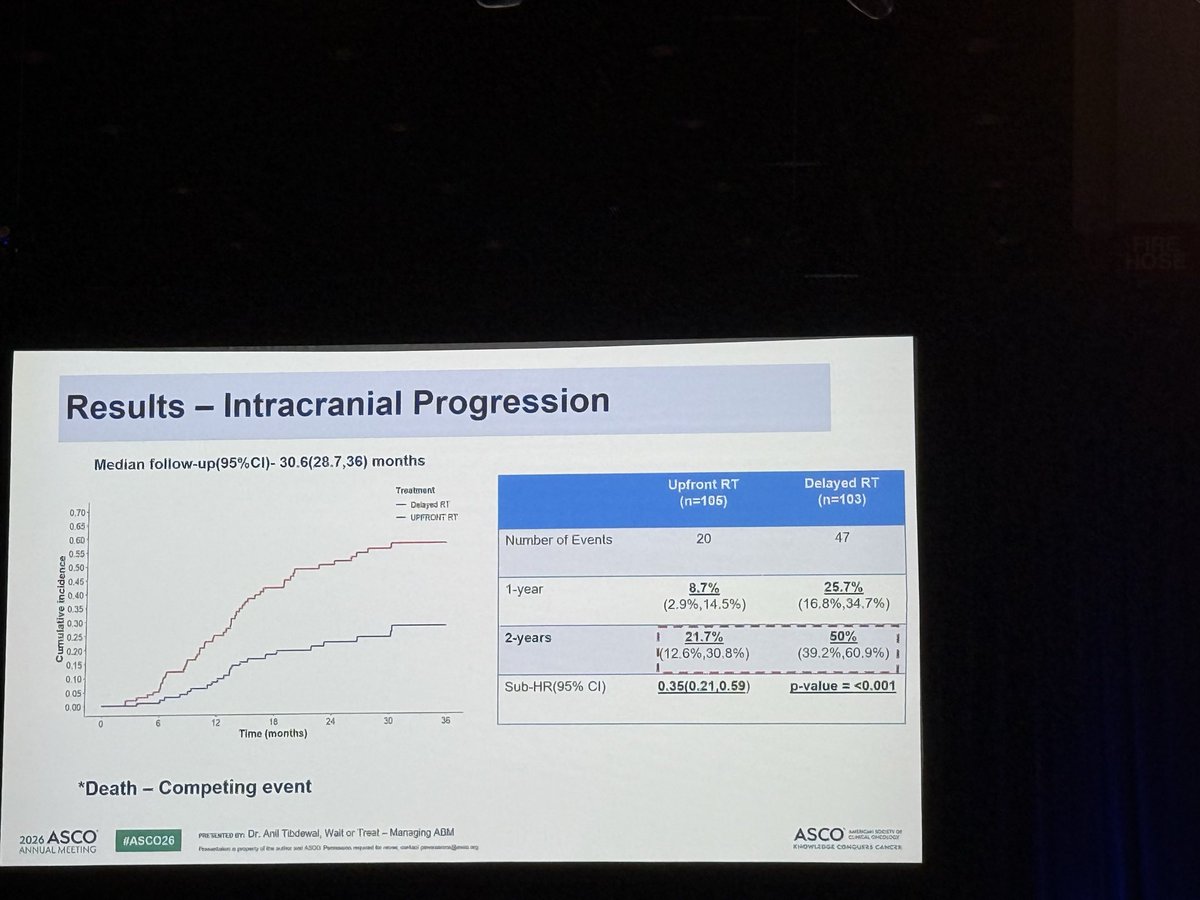
95% CI 0.21-0.59, p<0.001; 2y icPD 21.7% vs 50%
TL;DRUpfront cranial RT cut intracranial progression (sub-HR 0.35, 0.21-0.59, p<0.001) but 2y OS favored delayed RT, 48% vs 60%.
The intracranial win is real (2y progression 21.7% vs 50%, sub-HR 0.35) yet does not convert: 2y OS 48% upfront vs 60% delayed, HR 1.45. With necrosis reported ~6% upfront vs none delayed, and RT dose/technique unstated in source, this moves the timing decision toward deferral with MRI q3m surveillance.
In treatment-naive EGFR or ALK-driven metastatic NSCLC with asymptomatic measurable brain mets starting a TKI, this supports deferring cranial RT with q3m MRI rather than treating reflexively; it says nothing about symptomatic mets, large or dominant lesions, or oncogene-negative disease.
This is a timing question, not an omission question: both arms got cranial RT, and deferring cost 2y intracranial progression of 50% vs 21.7% while sparing necrosis (~6% upfront vs none delayed). Dose, fractionation, and WBRT-vs-SRS are absent from source, which is what gates transferring the toxicity read to your own technique.
The delayed arm's 1y intracranial progression of 25.7% on TKI plus chemotherapy is the number that makes upfront RT deferrable: most asymptomatic brain mets did not declare themselves in the first year. Deferral is contingent on q3m MRI surveillance, not on the TKI alone.
+2 more figures


10 details
Phase III open-label RCT, single-centre (Tata Memorial, Mumbai), N=208 randomised 1:1 to upfront (n=105) vs delayed cranial RT, both on TKI plus chemotherapy. Stratified by GPA (0-2 vs >2) and synchronous vs metachronous BM. Median follow-up 30.6 mo (28.7, 36).
Metastatic NSCLC with an EGFR or ALK alteration, ECOG PS 0-2, radiologically measurable brain metastases that were completely asymptomatic. Number, size, and location of lesions are not reported in source.
Upfront arm received cranial RT at diagnosis; the delayed arm received it at intracranial progression or patient's wish, so both arms are RT-exposed and the question is timing, not omission. Dose, fractionation, and technique (WBRT vs SRS) are not reported in source, which is the main barrier to transferring this result.
Primary: intracranial PFS. Secondary: OS, PFS, toxicity, ORR, neurocognition, PROM. Surveillance was MRI brain q3m for the first year, then q6m, which is what makes a delayed strategy safe to run.
Primary endpoint met. Survival ran the other way: 2y OS 48% upfront vs 60% delayed, OS HR 1.45, reported by attendees rather than captured in the slide OCR.
| Timepoint | Upfront RT (n=105) | Delayed RT (n=103) |
|---|---|---|
| 1-year | 8.7% (2.9%, 14.5%) | 25.7% (16.8%, 34.7%) |
| 2-years | 21.7% (12.6%, 30.8%) | 50% (39.2%, 60.9%) |
| Sub-HR (95% CI) | 0.35 (0.21, 0.59), p<0.001 | ref |
Radiation necrosis ~6% in the upfront arm and none in the delayed arm per attendee reports, and described as less severe when delayed. Full toxicity tables, neurocognition, and PROM were secondary endpoints not reported in the source.
The intracranial magnitude sits alongside the older WBRT-era data (QUARTZ, and the historic SRS-vs-WBRT trials) in showing that cranial RT controls the brain without buying survival. What is new is testing it where a CNS-penetrant TKI is the competing intracranial therapy, a setting those trials predate entirely.
Single-centre and open-label, and the RT prescription is absent from the source, so a reader cannot tell whether the necrosis signal reflects WBRT, SRS technique, or concurrent TKI exposure. Median follow-up of 30.6 mo is short relative to expected survival in this population, and the OS comparison was a secondary endpoint, not powered.
The result splits the two things RT is usually credited with: it clearly buys intracranial control, and it clearly does not buy time. Whether the OS direction is a real cost of upfront RT or noise in an underpowered secondary is the open question, and it decides whether deferral is merely non-inferior or actually preferred.
CONSORT flow
Randomised phase 3, prespecified intracranial PFS met, but the survival signal runs opposite the intracranial win. Directly contests reflex upfront cranial RT.
- Does the OS direction hold with longer follow-up?
- Was cranial RT whole-brain or stereotactic, at what dose?
- Neurocognition and PROM outcomes by RT timing
📚 Sources · 🐦 3 tweets
#ASCO26 | Wait or Treat? Brain RT in EGFR/ALK+ NSCLC
— OncLive.com (@OncLive) May 29, 2026
Presented by Dr Anil Ramakant Tibdewal.
A landmark Phase III randomized trial from @TataMemorial addressed a long-standing question: should asymptomatic brain metastases in oncogene-driven NSCLC receive upfront cranial RT or… pic.twitter.com/lRy9CfyQ8r
Should asymptomatic brain mets await systemic response in front line within EGFR/ALK context? I think yes. Despite reducing icPD, delayed brain RT OS looked better and radiation necrosis didn’t occur vs 6% #ASCO26 pic.twitter.com/O6d7GrvtU4
— Dr Riyaz Shah (@DrRiyazShah) May 29, 2026
No improvement in survival with up front radiation. OS favored delayed radiation with 2y OS 48% with early radiation vs 60% in late (OS HR 1.45). Also, radiation necrosis less common and less severe in delayed arm. Each case unique but delayed approach appealing #ASCO26 pic.twitter.com/wIhjqxhSaq
— Stephen V Liu, MD (@StephenVLiu) May 29, 2026